Biting Off What They Can Chew: Scientists Use 3D Reconstructions to Study Animal Jaw Mechanics


Studying the muscles that animals use to bite and chew can tell us a lot about their eating habits. In the past, researchers often undertook painstaking dissection of animal specimens by hand to visualize the muscles used for specific tasks like chewing. Physical damage from the separation of muscle layers during dissection and the effects of dehydration post-mortem can limit the accuracy of manual dissection, making this option less than ideal. In two recent PLOS ONE studies, researchers show us that modern “digital dissection” technologies can be used to avoid the problems associated with traditional dissections in two very different animals, one cute and cuddly and one slimy and giant.
In the first study, the authors used non-destructive X-ray computed tomography (CT) and magnetic resonance imaging (MRI) techniques alongside traditional dissection to generate the first detailed model of the muscles a common wombat uses to close its jaw. While we are most familiar with these types of scans from their use in medical imaging and screening for human diseases, these same methods can be used by researchers to get a peek at an animal’s inner muscular workings.
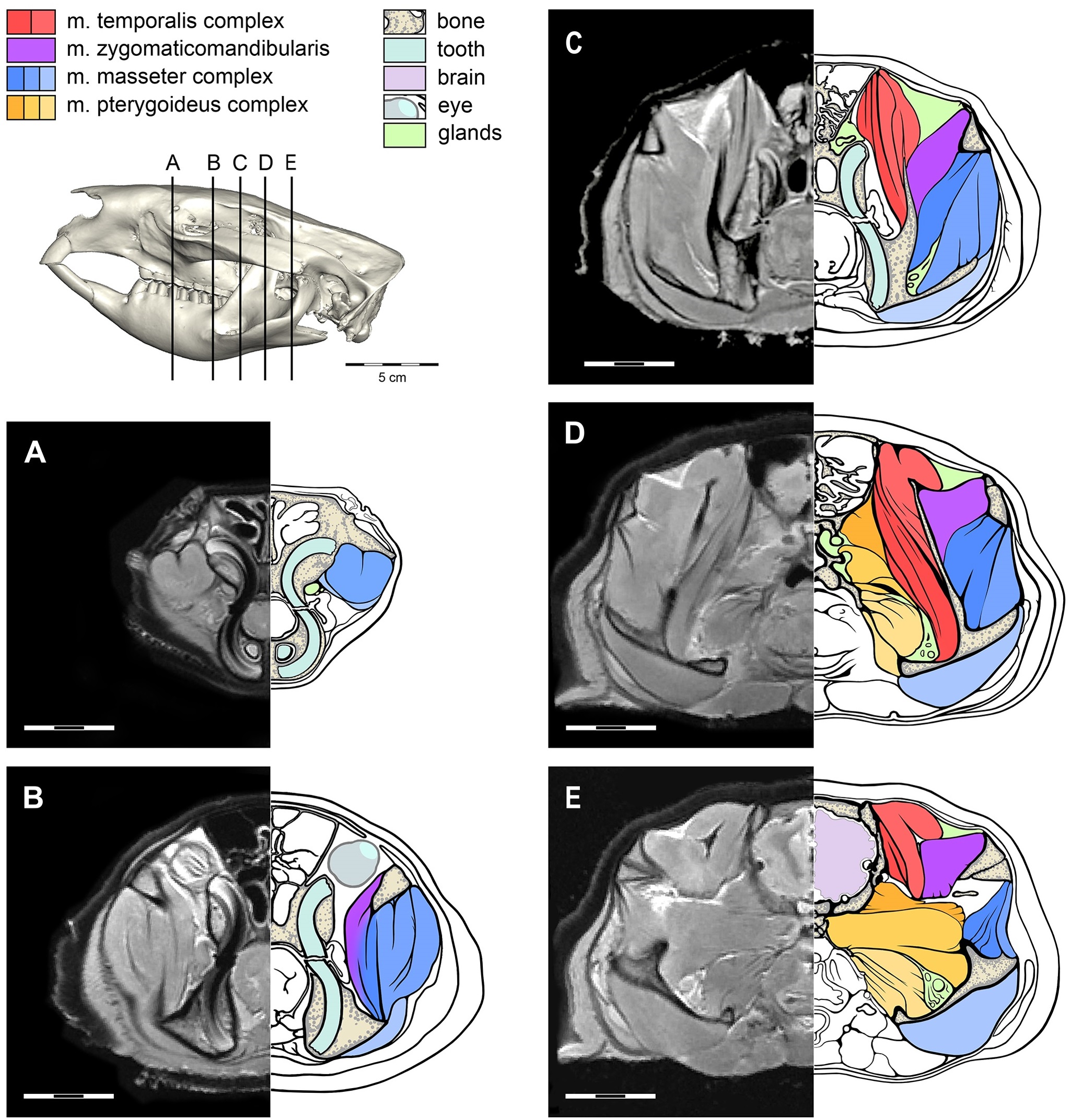
CT and MRI scans can pinpoint specific regions of an object and create a series of bisecting images, or “slices.” Scientists can view each slice separately, allowing them to identify individual muscle groups and tissues, as in the image below.
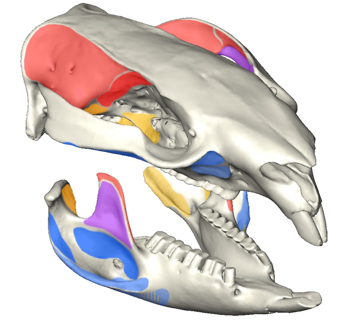
Individual slices are assembled to reconstruct the object with a detailed view of the inner structures. In the study, a 3D reconstruction of the wombat head is created by combining the information obtained from CT and MRI slices. Because these techniques allow the authors to see the inner architecture of the head, they are able to reconstruct the bones and muscles from the inside out, and see first-hand the interactions of the muscles in their position in the skull. What’s more, a neat, downloadable 3D PDF (Figure S1) is included in the paper that, when opened using Adobe Reader, is fully interactive. Readers can rotate the model 360°, isolate individual parts, or make certain parts transparent.

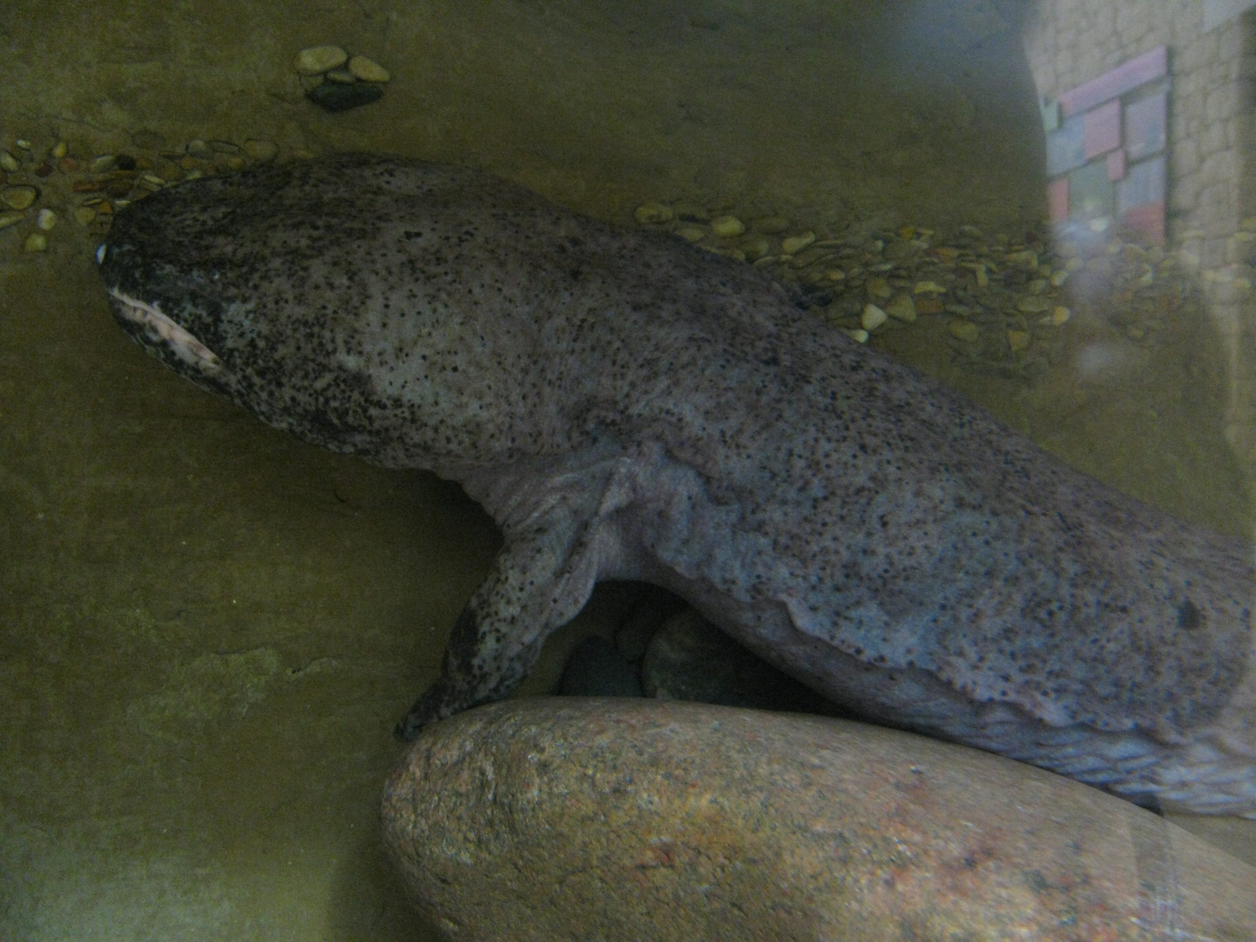
Authors of another recent PLOS ONE paper took their analysis one step further to study the bite mechanics of a Chinese giant salamander, the world’s largest amphibian. These researchers used CT scanning to analyze sutures that naturally occur on the skulls of adult and adolescent salamanders. The sutures are essential for skull stability and help to disperse compression forces during biting. By modeling the stress points that would occur under different biting conditions and comparing them to the skull sutures, scientists can predict which bite pattern would cause the least stress on the skull.
The authors of this study used computer simulations to model a bilateral bite, with initial equal pressure on both sides of the mouth, or a unilateral bite, with initial pressure on the left side of the mouth only. In each situation, authors simulated prey capture with the initial bite occurring toward the front versus the back of the mouth. The scientists found a clear difference in the simulated pattern of generated forces when the salamander was biting down at the front tip of the mouth versus the back.
In the video above, the authors used heat mapping to show the simulated forces exerted on various points in the salamander’s skull during the bite, with red patches indicating the greatest force, and blue patches indicating the least. The video shows what the researchers predict to be the optimal biting strategy: biting down at the front of the mouth causes less stress on the skull relative to biting down further back in the jaw.
While a bilateral bite distributes forces and allows salamanders to capture elusive or large prey, the video below shows an asymmetrical bite that may be utilized during a “sit-and-wait” strategy to capture prey in the water using a sudden strike on one side of the mouth. This asymmetrical strike is unique among vertebrates and can create a suction, pulling the prey and surrounding water into the mouth.
Both of these “digital dissection” techniques allow scientists to look at jaw mechanics in exquisite detail, without destroying the specimen, and may even help us understand the bites of extinct species. And maybe the next time you bite into a sandwich, you’ll think a whole lot more about just what goes into that chomp.
Citations:
Sharp AC, Trusler PW (2015) Morphology of the Jaw-Closing Musculature in the Common Wombat (Vombatus ursinus) Using Digital Dissection and Magnetic Resonance Imaging. PLoS ONE 10(2): e0117730. doi: 10.1371/journal.pone.0117730
Fortuny J, Marcé-Nogué J, Heiss E, Sanchez M, Gil L, et al. (2015) 3D Bite Modeling and Feeding Mechanics of the Largest Living Amphibian, the Chinese Giant Salamander Andrias davidianus (Amphibia:Urodela). PLoS ONE 10(4): e0121885. doi:10.1371/journal.pone.0121885
Images:
Image 1: Phil Whitehouse, Wombat via flickr
Image 2: Toby Jungen, Chinese Giant Salamander via flickr
Image 3, 5, and 6: 10.1371/journal.pone.0117730
Image 4: 10.1371/journal.pone.0121885
Videos: 10.1371/journal.pone.0121885